Introduction: Why Chest X-Rays Matter for Senior Health
As we age, routine diagnostic imaging becomes an essential tool for maintaining health and catching potential problems early. Chest X-rays, also called chest radiography, represent one of the most valuable screening and diagnostic tools available to seniors. According to the American Lung Association, chest radiography is often the first imaging test healthcare providers order when evaluating respiratory symptoms, heart conditions, or unexplained health changes in older adults.
“For seniors, chest X-rays serve as a window into multiple organ systems simultaneously,” explains Dr. Michael Curley, a board-certified family medicine and geriatric medicine physician with PromiseCare Medical Group who has provided care in the Inland Empire for over 37 years. “A single chest X-ray can reveal important information about the lungs, heart, blood vessels, ribs, and spine. This makes it an invaluable screening tool for the age-related conditions that commonly affect older adults.”
Understanding what chest X-rays can detect helps seniors make informed decisions about their health care and recognize when imaging might be beneficial. This comprehensive guide explores the diagnostic capabilities of chest radiography for older adults, when imaging is recommended, and what your physicians are looking for when they review your films.
How Chest X-Rays Work: The Basics of Diagnostic Imaging
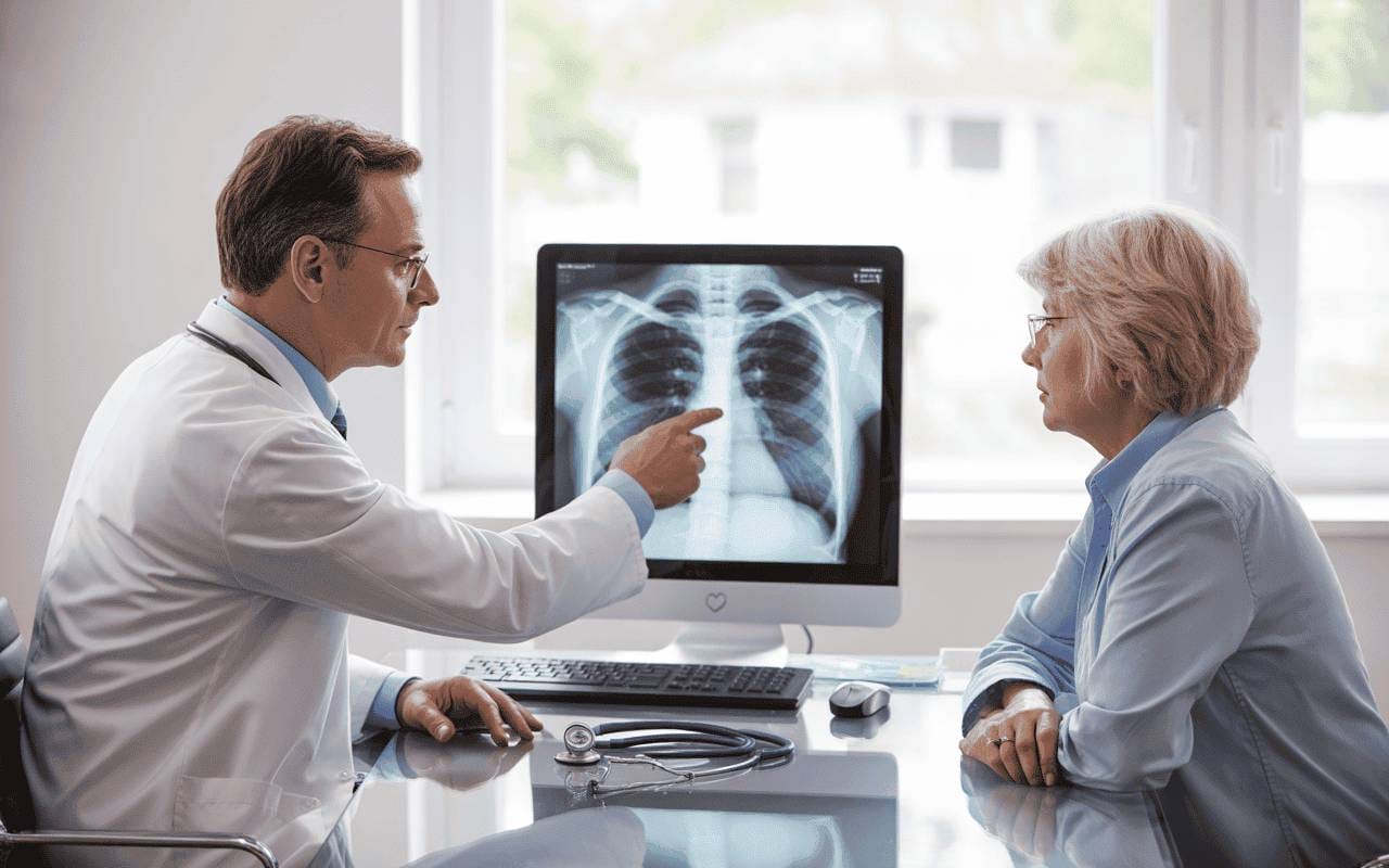
Chest radiography uses focused beams of radiation to create detailed images of structures inside the chest cavity. The process is quick, non-invasive, and painless, typically taking less than 15 minutes from start to finish. During the examination, X-ray beams pass through your body at different rates depending on tissue density, creating contrast that allows physicians to visualize organs, bones, and abnormalities.
Different tissues appear distinct on chest X-rays. Dense structures like bones and calcified areas appear white, soft tissues show up as varying shades of gray, and air-filled spaces like healthy lungs appear darker. This variation in appearance allows radiologists and physicians to identify normal anatomy and detect abnormalities.
Most chest X-rays involve two standard views: a posterior-anterior (PA) projection taken from behind and a lateral view from the side. These two perspectives provide comprehensive visualization of the chest structures and help physicians identify the precise location of any concerning findings.
What Chest X-Rays Can Detect in Seniors: A Complete Overview
Respiratory Conditions and Lung Disease
Pneumonia: Detecting Lung Infections Early
Pneumonia remains one of the leading causes of hospitalization and mortality in seniors. Chest radiography plays a critical role in diagnosing pneumonia by revealing areas of consolidation, infiltrates, or opacity in lung tissue that indicate infection.
“Seniors are particularly vulnerable to pneumonia due to age-related changes in immune function and the presence of chronic conditions,” notes Dr. Edivina Gonzales, an internal medicine physician with PromiseCare Medical Group. “Chest X-rays help us detect pneumonia early, determine its severity, and monitor response to antibiotic treatment. In elderly patients, we may see pneumonia on X-ray even before symptoms become severe.”
Chest radiography can identify:
- Community-acquired pneumonia presenting as patchy infiltrates
- Aspiration pneumonia often appearing in lower lung zones
- Bacterial vs. viral patterns helping guide antibiotic decisions
- Pleural effusion indicating fluid accumulation around lungs
- Complications such as abscesses or empyema
According to research published in the National Center for Biotechnology Information, chest X-rays are effective for early detection of respiratory infections, especially in older patients who may present with atypical symptoms.
Chronic Obstructive Pulmonary Disease (COPD) and Emphysema
While spirometry remains the gold standard for diagnosing chronic obstructive pulmonary disease, chest radiography provides valuable complementary information about lung structure and disease progression. Chest X-rays can reveal characteristic signs of COPD and emphysema that help physicians understand disease severity.
Key findings on chest X-rays in COPD patients include:
- Hyperinflation with flattened diaphragms
- Increased retrosternal airspace indicating trapped air
- Bullae formation showing large air-filled spaces
- Diminished vascular markings in affected lung areas
- Barrel chest appearance on lateral views
“When evaluating seniors for breathing difficulties, chest X-rays help us distinguish between COPD, heart failure, and other causes of dyspnea,” explains Dr. John Schoonmaker, a family practice and geriatric medicine specialist with PromiseCare Medical Group. “The X-ray patterns, combined with pulmonary function tests and clinical examination, guide our treatment approach.”
Research indicates that approximately 14% of chest X-rays performed at initial COPD assessment detect other potentially treatable conditions causing shortness of breath, making routine imaging valuable for comprehensive evaluation.
Lung Cancer: Early Detection Saves Lives
Chest radiography can identify suspicious lung nodules, masses, and tumors, though it’s not as sensitive as low-dose computed tomography (CT) for early-stage lung cancer screening. However, chest X-rays remain widely used due to accessibility, low cost, and minimal radiation exposure.
Chest X-rays may reveal:
- Pulmonary nodules appearing as rounded opacities
- Masses larger than 3 centimeters
- Hilar lymph node enlargement suggesting spread
- Pleural effusion which may indicate advanced disease
- Atelectasis from airway obstruction
The American Lung Association recommends annual low-dose CT screening for high-risk individuals, but chest X-rays often serve as the initial investigation when lung cancer is suspected based on symptoms like persistent cough, hemoptysis, or unexplained weight loss.
Pulmonary Fibrosis and Interstitial Lung Disease
Age-related changes in lung tissue can lead to scarring and stiffness conditions collectively called interstitial lung disease. Chest radiography can identify patterns suggestive of pulmonary fibrosis, including:
- Reticular patterns showing interstitial thickening
- Honeycombing in advanced fibrosis
- Ground-glass opacities indicating active inflammation
- Reduced lung volumes from decreased compliance
“Interstitial lung abnormalities are increasingly recognized in elderly patients, even those without diagnosed lung disease,” notes Dr. Gonzales. “When we see these patterns on chest X-rays, we can initiate appropriate monitoring and treatment to slow disease progression.”
Cardiovascular Conditions Visible on Chest X-Rays
Congestive Heart Failure and Pulmonary Edema
Chest radiography excels at detecting signs of congestive heart failure, a condition affecting approximately 6 million American adults. When the heart cannot pump effectively, fluid backs up into the pulmonary circulation, creating characteristic findings on chest X-rays.
Dr. Ratan Tiwari, a board-certified cardiologist with over 20 years of experience at PromiseCare Medical Group’s Healthy Heart Medical Center, explains the progression: “Heart failure manifests on chest X-rays in predictable stages. We look for vascular redistribution, Kerley B lines indicating interstitial edema, alveolar filling patterns, and pleural effusions. These findings help us assess severity and guide treatment intensity.”
Key cardiac findings include:
- Cardiomegaly with cardiothoracic ratio exceeding 50%
- Pulmonary vascular congestion with upper lobe blood vessel prominence
- Kerley B lines representing fluid in interlobular septa
- Bat-wing pattern of alveolar edema in acute failure
- Bilateral pleural effusions from fluid accumulation
Chest radiography helps distinguish cardiogenic pulmonary edema from other causes of respiratory distress. The vascular pedicle width, cardiac silhouette, and distribution of edema provide clues about whether fluid overload stems from cardiac dysfunction or other mechanisms like capillary permeability changes.
“In acute decompensated heart failure, chest X-rays guide immediate treatment decisions,” Dr. Tiwari adds. “They help us determine whether a senior needs diuretic therapy, respiratory support, or hospital admission. Follow-up X-rays show whether treatment is effectively clearing pulmonary congestion.”
Aortic Calcification and Cardiovascular Risk
Chest X-rays can reveal calcification in the aortic arch and aortic valve, providing valuable information about cardiovascular health and risk. Aortic calcification appears as white, dense areas along the aortic contour, particularly visible on lateral chest radiographs.
Research published in Atherosclerosis demonstrates that aortic arch calcification detectable on chest X-rays is a strong independent predictor of cardiovascular events, including myocardial infarction and stroke. The extent of calcification, graded from 0 to 3, correlates with age, blood pressure, diabetes status, and kidney function.
“When we see significant aortic calcification on routine chest X-rays, it alerts us to increased cardiovascular risk,” explains Dr. Curley. “This finding prompts us to optimize blood pressure control, manage cholesterol aggressively, and screen for coronary artery disease. It’s a simple but powerful risk marker readily available from standard imaging.”
Bone Health Assessment
Osteoporosis and Compression Fractures
While dedicated bone density testing remains the standard for diagnosing osteoporosis, chest X-rays provide important clues about bone health in seniors. Radiologists assess bone mineral density by evaluating rib and spine opacity. Thinned, osteopenic ribs may be difficult to visualize, suggesting low bone density.
More importantly, chest radiography can identify vertebral compression fractures, which affect more than one-third of seniors with osteoporosis. These fractures often occur without significant trauma and may cause:
- Height loss from vertebral collapse
- Kyphosis creating a hunched posture
- Back pain though some fractures are painless
- Reduced lung capacity from chest cavity compression
“Undiagnosed vertebral fractures are common in older adults,” notes Dr. Schoonmaker. “When chest X-rays reveal compression fractures, we initiate osteoporosis treatment to prevent future fractures and address pain management. We also investigate for causes of secondary osteoporosis like vitamin D deficiency or hyperparathyroidism.”
Research indicates that COPD patients frequently show both reduced bone mineral density and vertebral fractures on imaging, highlighting the importance of comprehensive evaluation.
Rib Fractures and Chest Wall Injuries
Chest radiography effectively identifies rib fractures from falls or trauma, conditions that become increasingly common with age-related balance problems and osteoporosis. While not all rib fractures require specific treatment, identifying them helps physicians:
- Provide adequate pain management
- Assess for underlying lung injury
- Monitor for delayed complications like pneumonia
- Determine if mobility assistance is needed during healing
Other Conditions Detected on Chest X-Rays
Tuberculosis Screening
For seniors entering long-term care facilities or those with risk factors for tuberculosis exposure, chest radiography plays an important screening role. Chest X-rays can identify:
- Active tuberculosis with characteristic upper lobe infiltrates and cavitation
- Latent TB with calcified granulomas (Ghon focus) from old infection
- Pleural involvement with effusion or thickening
- Post-TB changes like fibrothorax in elderly patients who had historical disease
“Tuberculosis screening is especially important in congregate living settings where transmission risk is higher,” explains Dr. Gonzales. “Public Health guidelines recommend chest X-rays for adults over 65 from high-risk groups before long-term care placement.”
Mediastinal Masses and Lymphadenopathy
The mediastinum, the central chest cavity containing the heart, great vessels, esophagus, and lymph nodes, can develop masses or enlarged lymph nodes that appear on chest X-rays. Abnormal mediastinal contours may indicate:
- Lymphoma or leukemia
- Metastatic cancer
- Thyroid masses
- Vascular abnormalities like aortic aneurysms
Sarcoidosis and Other Systemic Diseases
Chest X-rays can reveal patterns of hilar lymphadenopathy and pulmonary infiltrates characteristic of sarcoidosis, a granulomatous disease that can present at any age. Other systemic conditions with chest manifestations include rheumatoid arthritis, lupus, and progressive systemic sclerosis.
Age-Related Changes on Chest X-Rays: What’s Normal vs. Concerning
Senile Emphysema and Hyperinflation
Not all findings on chest X-rays in seniors indicate disease. Age-related changes called “senile emphysema” or “senile hyperinflation” reflect normal loss of lung elasticity with aging. These changes include:
- Enlargement of distal airspaces
- Some loss of supporting tissue
- Mild hyperinflation without significant bullae
“It’s important to distinguish normal aging changes from pathologic emphysema,” notes Dr. Curley. “Senile hyperinflation doesn’t cause symptoms and doesn’t progress like COPD-related emphysema. We use clinical correlation—smoking history, pulmonary function tests, and symptom assessment—to make this distinction.”
Tracheal Changes
Elderly patients may show tracheal deformities on chest X-rays, particularly the “saber-sheath” trachea characterized by narrowing of the coronal diameter. This finding is strongly associated with chronic obstructive pulmonary disease and affects predominantly male patients.
Calcifications
Multiple types of calcification become more common with age:
- Costal cartilage calcification along rib margins
- Tracheobronchial calcification in airway walls
- Granulomas from remote infections
- Pleural plaques from asbestos exposure or old inflammation
Most calcifications are benign and don’t require treatment, though they provide historical information about exposures and healed conditions.

When Seniors Should Get Chest X-Rays
Symptom-Based Indications
Chest radiography is indicated when seniors experience:
- Persistent cough lasting more than three weeks
- Shortness of breath new or worsening
- Chest pain unexplained by musculoskeletal causes
- Hemoptysis coughing blood
- Unexplained weight loss raising cancer concern
- Fever and respiratory symptoms suggesting pneumonia
- New heart failure symptoms including leg swelling and orthopnea
“We have a low threshold for ordering chest X-rays in seniors with respiratory or cardiac symptoms,” explains Dr. Tiwari. “The risk of serious underlying conditions increases with age, and chest radiography provides crucial diagnostic information quickly and non-invasively.”
Screening Scenarios
Beyond symptom evaluation, chest X-rays serve screening purposes in specific situations:
- Pre-operative assessment before surgery requiring general anesthesia
- Long-term care placement to screen for tuberculosis in high-risk populations
- Annual wellness visits for seniors with chronic heart or lung disease
- Post-hospitalization follow-up to ensure pneumonia has resolved
- Cardiac device implantation to verify proper lead placement
Lung Cancer Screening Considerations
For seniors at high risk of lung cancer—generally those aged 50-80 with a 20+ pack-year smoking history—low-dose computed tomography (CT) scanning provides superior early detection compared to chest X-rays. However, chest radiography may be appropriate for:
- Seniors who cannot undergo CT due to medical constraints
- Initial evaluation when lung cancer symptoms develop
- Monitoring known lung nodules at regular intervals
- Settings where CT access is limited
“While CT scanning has largely replaced chest X-rays for lung cancer screening, radiography remains valuable for diagnostic evaluation and follow-up,” notes Dr. Gonzales. “We tailor our imaging approach to individual patient circumstances, balancing detection capability with cost, accessibility, and radiation exposure.”
What to Expect During a Chest X-Ray Exam
Preparation and Procedure
Chest X-ray examinations require minimal preparation. Seniors should:
- Wear comfortable, loose-fitting clothing without metal fasteners
- Remove jewelry, eyeglasses, and other metal objects
- Inform the technologist about pacemakers or other implanted devices
- Mention any mobility limitations or positioning difficulties
For seniors with limited mobility, X-ray technologists can accommodate special positioning needs. Many facilities offer:
- Portable X-ray equipment for bedbound patients
- Chair supports for those who cannot stand
- Additional padding for comfort during positioning
- Extra time and assistance for seniors with dementia or confusion
The actual imaging takes only seconds. The technologist positions you in front of the X-ray plate, typically standing or sitting, then steps behind a protective barrier while capturing images. You’ll be asked to take a deep breath and hold it briefly—usually less than 10 seconds—while the X-ray is taken.
Radiation Safety in Seniors
Modern chest X-rays use minimal radiation, typically equivalent to 2-3 days of natural background radiation exposure. For seniors, the benefits of diagnostic information far outweigh the negligible cancer risk from low-dose radiation.
“Radiation concerns shouldn’t prevent necessary imaging in older adults,” reassures Dr. Curley. “The absolute risk of radiation-induced cancer decreases with age, while the immediate diagnostic value of chest X-rays remains high. We’re judicious about imaging frequency, but when clinically indicated, chest radiography is safe and appropriate for seniors.”
Results Timeline and Follow-Up
Most chest X-ray results are available within 24-48 hours. A radiologist reviews the images and provides a detailed interpretation to your physician, who then discusses findings and next steps with you.
If chest X-rays reveal concerning findings, your physician may recommend:
- Additional imaging with CT scans for detailed evaluation
- Pulmonary function testing to assess breathing capacity
- Echocardiography for heart function evaluation
- Blood tests to investigate infection or other conditions
- Specialist referral to pulmonology or cardiology
- Biopsy if masses or suspicious nodules are identified
Limitations of Chest X-Rays: When More Advanced Imaging Is Needed
While chest radiography provides extensive diagnostic information, it has limitations compared to more advanced imaging techniques.
Computed Tomography (CT) Superiority
CT scanning offers several advantages over chest X-rays:
- Early cancer detection identifying smaller nodules invisible on X-rays
- Three-dimensional visualization showing exact lesion locations
- Better soft tissue resolution for mediastinal masses
- Pulmonary embolism detection which chest X-rays cannot diagnose
- Quantitative assessment of emphysema extent and air trapping
“CT has revolutionized chest imaging, providing detail far beyond conventional X-rays,” explains Dr. Tiwari. “When chest X-rays show abnormalities, CT often becomes the next step for precise characterization. For lung cancer screening, pulmonary embolism evaluation, and detailed emphysema assessment, CT is indispensable.”
Echocardiography for Cardiac Assessment
While chest X-rays show cardiac size and pulmonary congestion, echocardiography provides functional information about:
- Heart valve abnormalities
- Ejection fraction and systolic function
- Diastolic function and filling pressures
- Regional wall motion abnormalities
- Pericardial effusion
Lung Ultrasound for Pulmonary Congestion
Bedside lung ultrasound has emerged as a valuable tool for assessing pulmonary congestion, showing B-lines that correlate with extravascular lung water. This point-of-care technique complements chest radiography in acute heart failure evaluation.
The Future of Chest Imaging for Seniors
Artificial Intelligence and Computer-Aided Detection
Advanced artificial intelligence algorithms are increasingly being applied to chest radiograph interpretation. These systems can:
- Detect subtle pneumonia infiltrates
- Identify small pulmonary nodules
- Flag tuberculosis patterns
- Quantify heart size automatically
- Prioritize worrisome findings for radiologist review
Research published in Frontiers in Big Data demonstrates that AI-based radiodiagnosis systems show promise in reducing radiologist workload while improving detection accuracy, particularly valuable given the billions of chest X-rays performed annually worldwide.
Digital Tomosynthesis
Digital tomosynthesis represents an evolution of conventional chest radiography, creating quasi-three-dimensional images from multiple X-ray exposures at different angles. This technique improves visualization of lung nodules and eliminates overlapping structures that can obscure pathology on standard films.
Mobile and Home-Based Imaging
For homebound seniors with mobility limitations, portable chest X-ray services bring diagnostic imaging directly to patients in their homes or long-term care facilities. Mobile radiologic technologists use compact equipment that produces diagnostic-quality images comparable to hospital-based examinations.
“Mobile X-ray services eliminate the stress and physical burden of transporting frail elderly patients to imaging centers,” notes Dr. Schoonmaker. “Medicare typically covers these services when patients are homebound due to severe illness, dementia, or mobility impairments. This innovation ensures seniors can receive necessary diagnostic evaluation safely in familiar surroundings.”
Frequently Asked Questions About Chest X-Rays for Seniors
How often should seniors get chest X-rays?
Chest X-ray frequency depends on individual health status and risk factors rather than age alone. Seniors with stable chronic conditions like COPD or heart failure may benefit from annual chest radiography to monitor disease progression. Those with acute symptoms require imaging as clinically indicated. There’s no routine screening schedule for asymptomatic seniors without risk factors.
Can chest X-rays detect all lung problems?
While chest radiography identifies many pulmonary conditions, it has limitations. Small lung nodules under 1 centimeter may be invisible. Early pneumonia can sometimes appear before X-ray changes develop. Pulmonary embolism and subtle interstitial lung disease may not show on standard films. When suspicion for serious lung pathology is high despite normal chest X-rays, CT scanning provides definitive evaluation.
What does an abnormal chest X-ray mean for seniors?
Not all abnormal chest X-ray findings indicate serious disease. Many elderly patients show benign age-related changes like calcifications, mild hyperinflation, or old granulomas from remote infections. Your physician correlates X-ray findings with symptoms, physical examination, and medical history to determine clinical significance and whether additional testing is warranted.
Are chest X-rays safe for seniors with multiple medical problems?
Chest radiography is one of the safest medical tests available. The minimal radiation exposure poses negligible risk at any age. Seniors with pacemakers, implanted defibrillators, or other medical devices can safely undergo chest X-rays. The examination requires no contrast dye injection and has virtually no contraindications. Even seniors with severe illness can typically tolerate the brief procedure.
How do physicians know if pneumonia on a chest X-ray is improving?
Follow-up chest X-rays, typically performed 4-6 weeks after pneumonia diagnosis, show whether infiltrates have cleared. However, radiographic improvement often lags behind clinical recovery. Seniors may feel better while X-ray abnormalities persist. Complete clearing can take several months, particularly in elderly patients with underlying lung disease or immune compromise.
Will chest X-rays detect coronavirus (COVID-19) in seniors?
Chest X-rays can identify pneumonia patterns suggestive of COVID-19, particularly in older patients who tend to show more pronounced radiographic changes than younger individuals. Research presented at the American Roentgen Ray Society found chest radiography more effective in diagnosing COVID-19 in people older than 50 years, with 93% showing abnormal findings. However, chest radiography supplements rather than replaces COVID-19 testing.
Can chest X-rays show if heart medications are working?
Chest radiography can demonstrate whether heart failure treatment is reducing pulmonary congestion. Sequential X-rays showing decreased cardiomegaly, clearing of Kerley B lines, and resolution of pleural effusions indicate improving cardiac function and effective diuretic therapy. However, echocardiography provides more precise assessment of cardiac function and ejection fraction changes.
What happens if chest X-rays show a suspicious lung nodule?
When chest radiography identifies a pulmonary nodule, your physician considers nodule characteristics (size, shape, density), your smoking history, age, and cancer risk factors. Most lung nodules in seniors are benign granulomas from old infections. However, nodules larger than 8mm, irregular shaped, or growing over time warrant further evaluation with chest CT scanning. Depending on CT findings, options include surveillance imaging, PET scanning, or biopsy.
Do all seniors need chest X-rays before surgery?
Routine pre-operative chest X-rays are not necessary for all elderly surgical patients. Current guidelines recommend pre-operative chest radiography for seniors undergoing major surgery, particularly thoracic or cardiac procedures, or when history and physical examination suggest undiagnosed heart or lung disease. Many seniors undergoing minor procedures don’t require routine chest imaging unless symptoms or chronic conditions warrant evaluation.
Can chest X-rays diagnose the cause of chronic cough in seniors?
Chest radiography helps identify structural lung abnormalities, tumors, infections, and heart failure that may cause chronic cough. However, many causes of persistent cough in seniors—including gastroesophageal reflux, post-nasal drip, medication side effects (particularly from ACE inhibitors), and asthma—produce normal chest X-rays. When cough persists despite normal radiography, additional evaluation including CT scanning, pulmonary function testing, and gastroenterology consultation may be necessary.

Conclusion: The Enduring Value of Chest Radiography in Senior Care
Chest X-rays remain an indispensable diagnostic tool in geriatric medicine, providing rapid, accessible, and cost-effective evaluation of cardiopulmonary health. From detecting pneumonia and heart failure to identifying lung cancer and bone fractures, chest radiography offers comprehensive diagnostic information that guides treatment decisions and improves outcomes for older adults.
“After nearly four decades practicing family medicine and geriatrics, I’m continually impressed by how much information a simple chest X-ray provides,” reflects Dr. Curley of PromiseCare Medical Group. “For seniors dealing with multiple chronic conditions, chest radiography offers a holistic assessment that can detect problems early when they’re most treatable. It’s a cornerstone of senior health maintenance.”
Understanding what chest X-rays can reveal empowers seniors and their families to appreciate the value of this common test and recognize when imaging might be beneficial. While newer technologies like CT scanning and lung ultrasound offer enhanced capabilities for specific indications, conventional chest radiography continues serving as the frontline imaging modality for evaluating respiratory and cardiac concerns in the elderly population.
PromiseCare Medical Group, the Inland Empire’s largest Independent Physician Association network, provides comprehensive senior care with access to advanced diagnostic imaging and specialist services across Riverside County. If you’re experiencing symptoms that might warrant chest X-ray evaluation, contact your primary care physician at PromiseCare to discuss whether imaging would be appropriate for your situation.
About PromiseCare Medical Group
PromiseCare Medical Group represents the longest continually serving and largest Independent Physician Association network in the Inland Empire region of California. With more than 60 primary care doctors and 400 specialists, PromiseCare places the needs of patients first, focusing on excellent clinical outcomes, patient safety, and exceptional service. The network includes teams of dedicated physicians, hospitals, diagnostic centers, and nursing staff throughout Riverside County, providing stability and consistency in serving every patient today and tomorrow.
Medical Disclaimer: This information is for educational purposes only and should not replace professional medical advice. Always consult with your physician regarding symptoms, diagnostic testing, and treatment recommendations appropriate for your specific health situation.