 Similar to visible light, X-rays produce electromagnetic radiation. X-rays, however, have a much higher energy than light and can travel through any object, including the human body. Images of tissues and structures inside the body can be generated using medical x-rays. X-rays traveling through the body will pass through a detector on the opposite side of the patient, which will produce an image representing “shadows” created by the objects in the body.
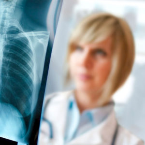
There are numerous types of x-ray detectors that are used to produce digital images, including photographic film. An x-ray image obtained from this process is a radiograph.

A radiograph is created by positioning the patient so that the part of the body to be imaged is situated between the x-ray source and the detector. The radiation from the machine travels through the body and is absorbed in different amounts by different tissues depending on their density. Both density and atomic number (the number of protons in an atom’s nucleus) determine the radiological density of materials being imaged.
Calcium, for instance, is an atomic number-higher substance than most tissues, which makes it suitable for structures like bone. This property makes bones readily absorb x-rays and produce high contrast on the x-ray detector as a result. The resulting contrast between the black background of a radiograph and bony structures makes them appear whiter than other tissues.
Radiation travels more easily through tissues that are less dense radiologically, such as fat and muscle, and through air-filled cavities. Radiographs show these structures as shades of gray.
It’s crucial to remain still while the images are taken. This ensures the best quality images.
On the X-ray table, the patient is placed between the X-ray machine and a cassette containing the X-ray film or specialized image plate, with the body part to be X-rayed carefully positioned between the machine and the cassette. In some cases, the patient may sit or stand while the examination is conducted.
An X-ray shield (apron) can be used to prevent the exposure of body parts not being imaged.
 The diagnostic benefits of x-ray scans outweigh the risks substantially when used appropriately. Diagnoses such as blocked blood vessels, bone cancer, and infections can be made using X-rays.
On the other hand, x-rays produce ionizing radiation, which can adversely affect living tissue. A person’s risk increases with the number of exposures they receive over the course of their lives. Exposure to radiation, however, poses a low risk of cancer development.
If the area of the body imaged isn’t the abdomen or pelvis, there are no known risks to the baby from an x-ray in a pregnant woman. Doctors generally prefer exams that do not use radiation, such as MRIs or ultrasounds, when imaging the abdomen or pelvis. X-rays are an acceptable alternative imaging option if neither of these provides the answers needed, or if an emergency arises or there is another time constraint.
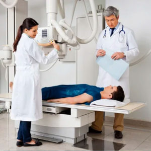 A radiology technician positions the patient on an x-ray table and places an x-ray film holder or recording plate under the table under the area to be imaged. To assist you in maintaining the proper position, sandbags, pillows, or other positioning devices may be used. To protect against radiation, a lead apron may be placed over your pelvic area or breasts.
When the technologist takes the x-ray, you may have to hold your breath for a few seconds while you are still. This helps to reduce the likelihood of a blurred image. The x-ray technician will walk behind a wall or into the next room in order to turn on the equipment.
The process may be repeated for another view. Typically, two or three images will be taken (from differing angles).
You may be asked to wait until the radiologist confirms they have all the images after the examination is complete.
 You can change back into your regular clothing after your X-ray images have been collected. If you are in good health, you may be able to go about your normal activities. You may have access to your results the same day as your procedure, or later.
After reviewing your X-rays and the radiologist’s report, your doctor will determine how to proceed. To develop an accurate diagnosis, they may order additional tests based on your results. These may include additional imaging scans, blood tests, or other diagnostic measures. The doctor may also prescribe treatment.
If you would like more information about your condition, diagnosis, or treatment options, ask your doctor.